A postoperative wound that fails to follow the expected trajectory is a diagnostic prompt, not a nuisance. The patient may look stable at the door and still have a collection under the fascia. The following composite case traces a familiar pattern: a wound that seemed minor at first, then declared itself with rising pain, discharge, and delayed healing. The value of the case lies less in a single clever intervention than in sequence—recognize, control the source, characterize the microbiology, choose a dressing that serves the wound, and follow the patient long enough to catch the next turn.
Recognition: the wound that stops improving
Infection after surgery rarely announces itself with a single dramatic sign. More often it accumulates: pain that increases rather than settles, erythema that extends beyond the incision, warmth, swelling, purulent or serosanguineous discharge, and a wound that stops contracting. Systemic features—low-grade fever, malaise, tachycardia—may be absent, especially in older or immunosuppressed patients. That is why serial examination matters. A wound that looked acceptable on Monday can be a different problem by Thursday.
The first task is to separate expected postoperative inflammation from infection. Early inflammation is usually confined to the incision, improves over days, and responds to elevation and simple analgesia. Infection tends to worsen, spreads, or produces discharge with odor or pus. Do not rely on a single photograph or a distant glance. Inspect the wound with adequate lighting, palpate gently, and measure the visible defect. Ask about pain out of proportion to the appearance, fever, and appetite. If the patient is systemically unwell or the wound is rapidly progressive, escalate immediately.
Source control comes before dressing choice
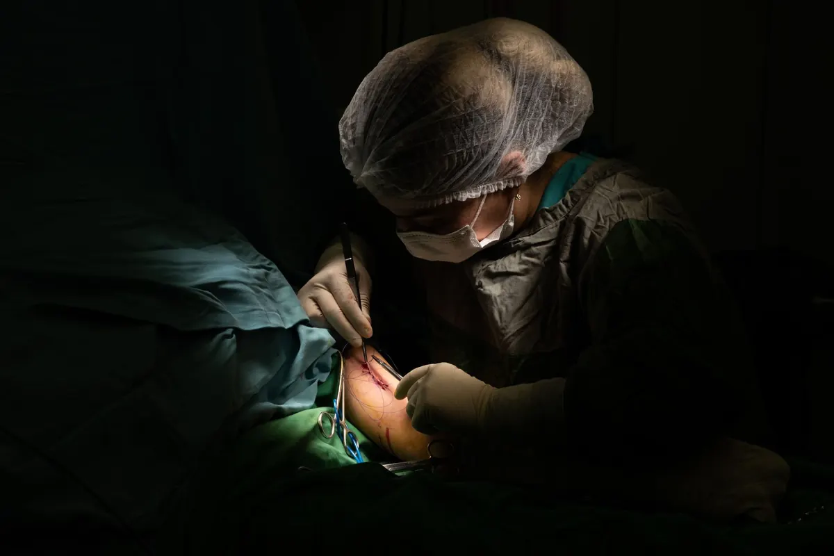
Source control is the pivot. A dressing cannot compensate for an undrained abscess, a retained foreign body, or necrotic tissue. In the case described, the wound edge looked clean but the cavity extended beneath the fascia. Gentle probing and imaging—guided by local practice—revealed a collection. The patient went to theatre for drainage and debridement.
The principles are consistent:
- Open and drain collections.
- Debride nonviable tissue until healthy bleeding tissue is reached.
- Remove foreign material—sutures, mesh, drains—when it is contributing and safe to do so.
- Irrigate with a solution appropriate to the tissue and local protocol; avoid cytotoxic agents that impair healing.
- Reassess the need for antibiotics after source control, not before.
Not every wound needs theatre. Superficial infections may respond to opening the skin, cleaning, and dressing. But deep, extensive, or systemic infection needs senior review. If in doubt, ask for help early.
Microbiology that changes management
Microbiology should answer a question: which organisms are present, and do they change treatment? A superficial swab from the wound surface is often unhelpful because it grows colonizers. A deep tissue sample, obtained during debridement or by aspiration of a collection, is more useful. Send tissue for Gram stain, culture, and sensitivity. If the patient is febrile or systemically unwell, blood cultures may be appropriate. Take cultures before starting or changing antibiotics whenever it is practical, but do not delay source control to wait for results.
In the case, cultures grew a mixed population of gram-positive cocci and gram-negative bacilli—a common pattern in postoperative wounds, especially after abdominal or perineal surgery. The result did not justify a broad-spectrum drug for every patient; it allowed the team to narrow therapy once the patient improved. Antibiotics are adjuncts to source control, not a substitute. Review them daily: is the patient improving, is the organism covered, can the spectrum be reduced, and is the duration appropriate? Local resistance patterns and hospital guidance should drive choices.
Dressing strategy: match the plan to the wound
Dressing selection follows assessment. The wound does not need a fashionable product; it needs a strategy matched to exudate, bioburden, dead space, and periwound skin. In the early inflammatory phase, frequent changes may be needed to monitor and clean. As the wound becomes cleaner, the interval can lengthen.
Ask four questions:
- Is exudate high, moderate, or low? Choose absorbency accordingly.
- Is there slough, eschar, or a cavity? Consider debridement, packing, or negative pressure in selected cases.
- Is bioburden controlled? Use an antimicrobial dressing only when indicated and for a defined period.
- Is the surrounding skin macerated or fragile? Protect it with a barrier film or appropriate dressing.
Negative pressure wound therapy can be useful in selected complex wounds after adequate debridement, but it is not a rescue for an undrained collection. For cavities, avoid packing too tightly; it can impede drainage and cause pressure. Document the dressing, the reason for it, and the planned review. If a dressing is not improving the wound within a reasonable interval, change the plan rather than repeating it.
Follow-up and the long tail of wound infection
Follow-up is where complex wounds are won or lost. The patient should leave with clear instructions: whom to call, what symptoms warrant urgent review, and when the next appointment is. At each visit, measure the wound, record exudate and odor, assess pain, and photograph with consent and local policy. Trends matter more than a single snapshot. A wound that is smaller but more painful, or cleaner but surrounded by spreading erythema, needs reassessment.
The case had a second turn: after discharge, the wound edge began to break down again. Because the patient had been given a scheduled review and a direct contact route, the team caught it early. A small recurrent collection was drained, and the dressing plan was simplified. That is the ordinary work of complex wound care—not a single heroic decision, but repeated small corrections.
Also address nutrition, glycaemic control, smoking, and pressure offloading where relevant. These factors influence healing, and patients often need practical help rather than a lecture. A dietitian, diabetes team, or tissue viability nurse can be valuable. Document who is responsible for what.
A practical checklist for the next complex wound
Before the next complex postoperative wound, use a short checklist:
- Confirm the wound is not hiding a deeper collection.
- Achieve source control before escalating dressings or antibiotics.
- Send deep tissue, not a superficial swab, when microbiology will change management.
- Match the dressing to exudate, dead space, bioburden, and skin.
- Schedule follow-up with measurements, photographs, and a clear escalation route.
Complex wound infection is rarely managed in one visit. It is managed in a sequence of good decisions, each reviewed against the patient in front of you. When the wound is deep, the patient is unwell, or progress stalls, seek senior surgical and specialist wound advice. General guidance cannot replace individual clinical judgment and local protocols.
Photo: Shutterbug26 / Pixabay